Immunofluorescence Protocol For Paraffin Embedded Sections

Protocol Guide Immunofluorescent Staining Of Whole Mount Organoids Using Antibodies Sigma Aldrich

Immunolabeling Molecular Life Science Learning Centers

Immunofluorescence Lab Tech Study Materials Study Time

2017 Fluomute Ready To Use Reagent To Reduce Autofluorescence In Cells And Tissue Just Incubate Fixed Cells Of Tissue Cells And Tissues Stem Cells Treatment

Staining For Ki67 In Paraffin Embedded Tissue Sections Nextgen Protocols

Anti Ki67 Antibody Ko Tested Ab15580 Abcam

April 10 2014 katie crosby 1 jessica simendinger 1 christopher grange 1 michelle ferrante 1 trisha bernier 1 claire standen 1.
Immunofluorescence protocol for paraffin embedded sections.

Help With High Background Staining With Immunofluorescence Of Tumors
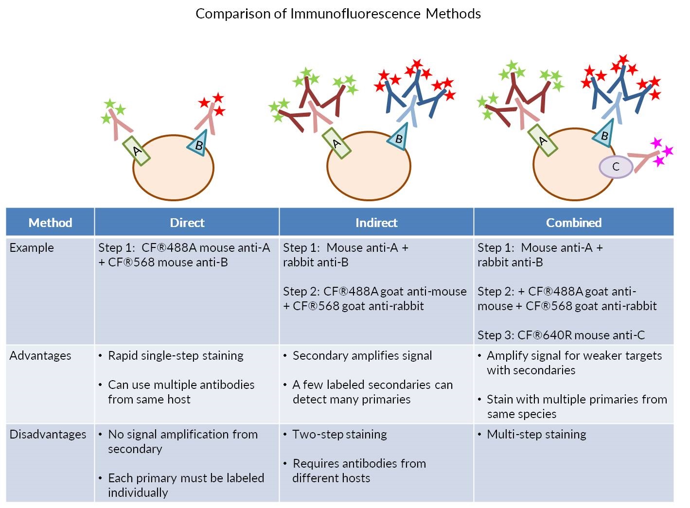
Considerations For Immunofluorescence Staining Biotium
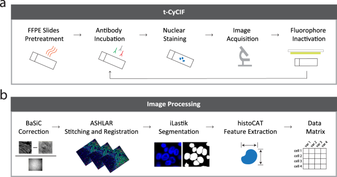
Highly Multiplexed Immunofluorescence Images And Single Cell Data Of Immune Markers In Tonsil And Lung Cancer Scientific Data

Anti Brdu Antibody Bu1 75 Icr1 Proliferation Marker Ab6326 Abcam

Recombinant Anti Cdx2 Antibody Epr2764y Ab76541 Abcam

Multilabel Immunofluorescence And Antigen Reprobing On Formalin Fixed Paraffin Embedded Sections Novel Applications For Precision Pathology Diagnosis Modern Pathology

Immunofluorescence Tissue Staining Youtube

Anticuerpos Antinucleares Ifi Con Patron Homogeneo Sobre Celulas Hep 2 Ana Convenience Store Products Natural Landmarks
Immunofluorescence Troubleshooting Protocol Tips Stressmarq

Recombinant Anti Sox9 Antibody Epr14335 78 Ab185966 Abcam

Recombinant Anti Irs2 Antibody Epr904 2 Ko Tested Ab134101 Abcam

Rnascope Multiplex Fluorescent Assay Advanced Cell Diagnostics Fluorescent Nebula Cell

Recombinant Anti Alpha Tubulin Antibody Ep1332y Microtubule Marker Ab52866

Recombinant Anti M6pr Cation Independent Antibody Epr6599 Ko Tested Ab124767

Recombinant Anti Kdel Antibody Epr12668 Ab176333 Abcam

Recombinant Anti Pax2 Antibody Ep3251 Ab79389 Abcam

Anti Vimentin Antibody V9 Cytoskeleton Marker Ko Tested Ab8069 Abcam

Recombinant Anti Syndecan 1 Antibody Epr6454 Ab128936 Abcam
1

Recombinant Anti Active Yap1 Antibody Epr19812 Ko Tested Ab205270 Abcam
What Is The Best Practice Staining On Slides Or Free Floating
Recombinant Anti Aquaporin 5 Antibody Epr3747 Ab92320 Abcam
Blog Immunofluorescence Vs Immunocytochemisty Vs Immunohistochemistry What Is The Difference

Recombinant Anti Notch1 Antibody Ep1238y Ko Tested Ab52627 Abcam
Source : pinterest.com
